Call Us Today:
561-861-1388
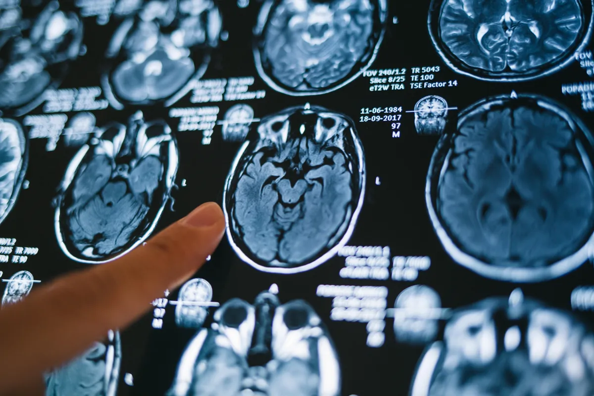
What is MRI?
What is MRI?
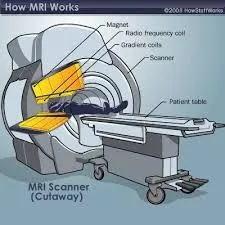
How does it work?
Magnetic Resonance Imaging (MRI) is a medical imaging technique used to visualize detailed internal structures of the body. The process of how an MRI machine works and how images are created can be explained in several steps:
Magnetic Field : An MRI machine contains a powerful magnet, usually a superconducting magnet. This creates a strong, uniform magnetic field around the patient. This magnetic field aligns the protons in the hydrogen atoms of the body's water molecules.
Radiofrequency Pulses : The machine emits radiofrequency (RF) pulses through a coil. These pulses are of a specific frequency and are directed at the body part being imaged. When the RF pulses are applied, they disturb the alignment of the hydrogen protons.
Resonance and Relaxation :
Excitation :
The RF pulses excite the protons, causing them to absorb energy and move out of alignment with the magnetic field.
Relaxation :
When the RF pulses are turned off, the protons gradually return to their original alignment. During this process, they release the absorbed energy.
Signal Detection : As the protons relax and return to alignment, they emit RF signals. These signals are detected by receiver coils in the MRI machine.
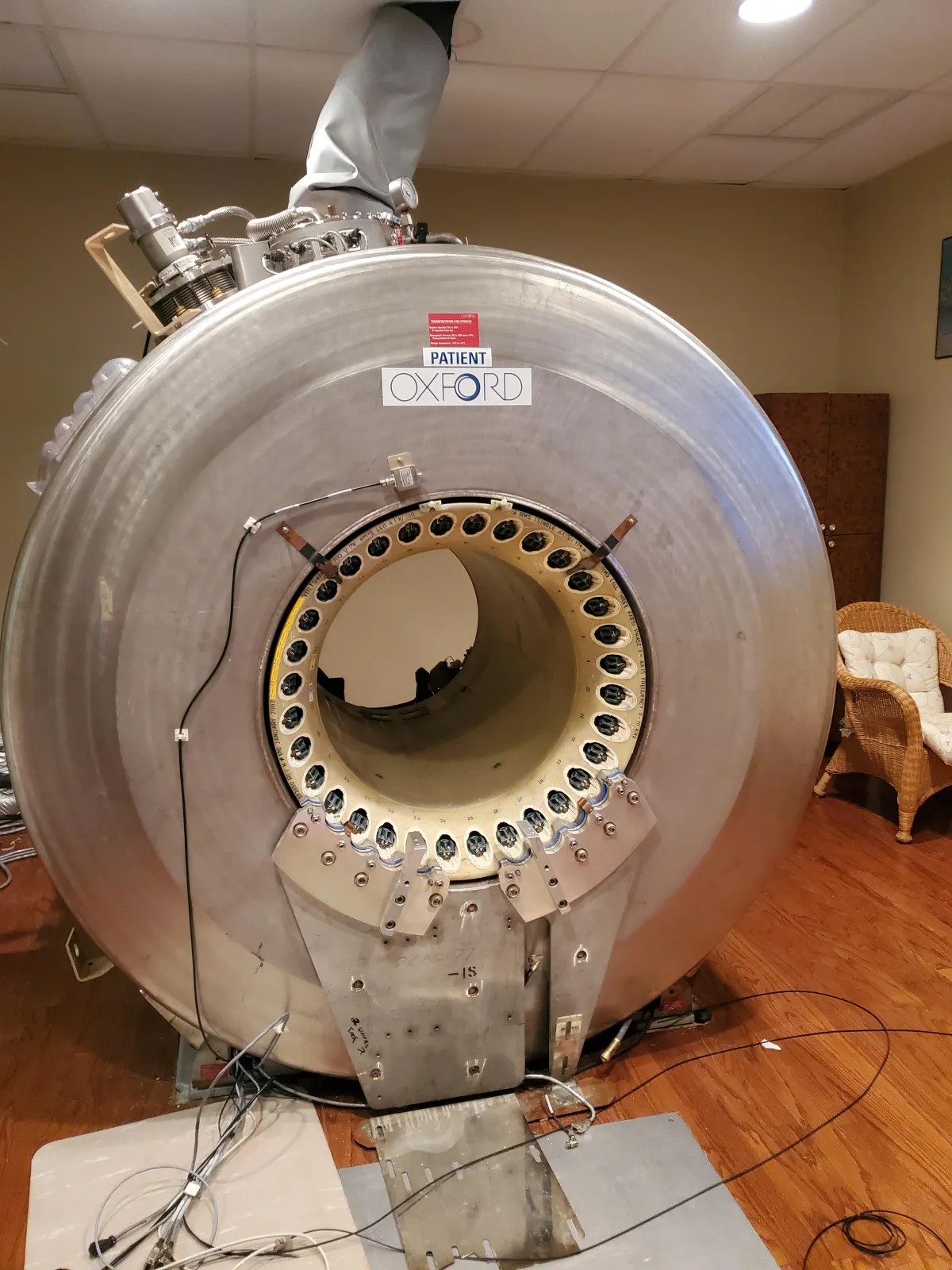
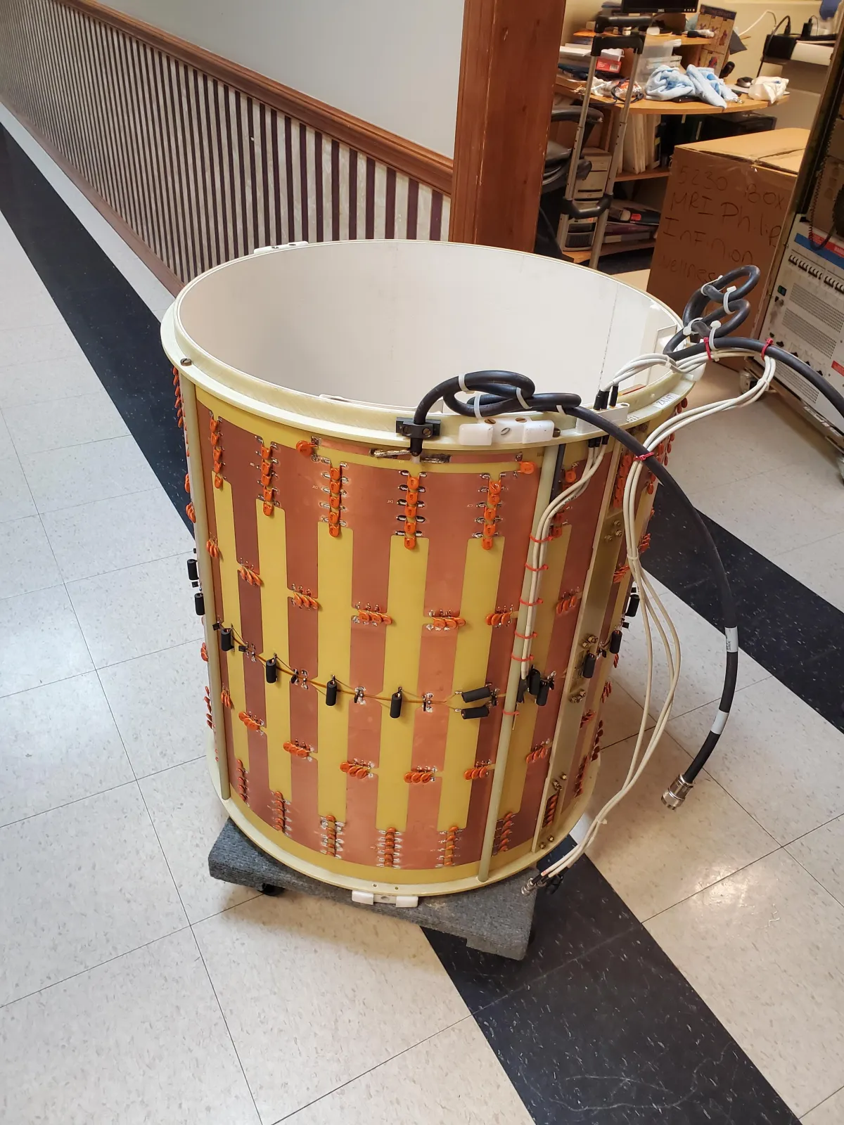
MRI RF COIL
Image Creation
Spatial Encoding : The MRI machine uses gradient magnets to vary the magnetic field in a controlled manner along different axes (X, Y, and Z). This spatial variation allows the machine to determine the
location of the signals.
Fourier Transformation : The received signals are complex and contain information about the location and type of tissues. A mathematical process called Fourier transformation converts these signals into a 2D or 3D image. The transformation separates the signals based on their frequency and phase, allowing the construction of detailed images.
Image Reconstruction : The MRI machine's computer processes the transformed data to create cross-sectional images (slices) of the body. These images can be stacked to form 3D representations of
the internal structures.
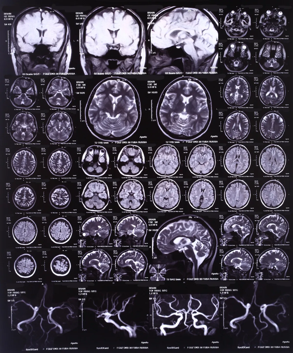
Types of MRI Sequences
Different MRI sequences (pulse sequences) can be used to highlight various tissues and abnormalities:
T1-Weighted Images : These provide high contrast
between different tissue types and are useful for visualizing anatomical details.
T2-Weighted Images : These are particularly good
for detecting abnormalities such as edema, tumors, and inflammation.
Diffusion-Weighted Imaging (DWI) : This sequence
is sensitive to the movement of water molecules and is useful in detecting stroke and other conditions affecting tissue microstructure.
Functional MRI (fMRI) : This measures brain
activity by detecting changes in blood flow, commonly used in neuroscience research.
Safety and Advantages
MRI is non-invasive and does not use ionizing radiation (unlike X-rays or CT scans), making it safer for repeated use. However, because it involves strong magnetic fields, it is essential to ensure that patients do not have any metal implants or devices that could be affected by the magnet.
In summary, MRI works by using strong magnetic fields and radiofrequency pulses to excite hydrogen protons in the body's tissues. The emitted signals are then detected, spatially encoded, and transformed into detailed images through complex computational processes.
© 2024 Wellness Associates of Florida, LLC